
How Scientific Photography AI Generation Shook Nikon Small World
The rapid rise of scientific photography AI generation has sent shockwaves through the global research community, forcing a reckoning over where empirical observation ends and digital fabrication begins. For fifteen years, the Nikon Small World In Motion competition has stood as the premier showcase for the invisible world. It is a space where researchers, pathologists, and hobbyists share breathtaking footage captured through the lenses of powerful microscopes. But the 2024 competition will be remembered not for a breakthrough in imaging, but for a high-profile disqualification that has exposed the vulnerabilities of scientific publishing and competition in the age of synthetic media.
Scientific photography AI generation refers to the use of artificial intelligence models to synthesize, alter, or fabricate microscopic and scientific imagery. While generative AI can enhance visualization, its unauthorized use in competitions like Nikon's Small World compromises empirical integrity, leading to disqualifications when synthetic data is presented as genuine empirical observation.<\/p>
- The Disqualification: Nikon stripped Dr. Ning Xu of his first-place prize in the Small World In Motion contest after confirming the video used generative AI.
- The Whistleblowers: Clinical experts immediately recognized that the depicted cellular structures were biologically impossible and lacked real-world noise.
- The Diagnostic Risk: Fabricated medical imagery compromises patient trust and complicates the training of diagnostic models for rare diseases like PCD.
- The Policy Shift: Nikon is actively re-evaluating its submission verification procedures to combat sophisticated microscopic image manipulation.
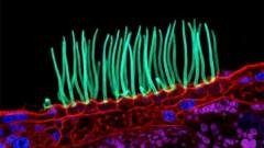
The controversy erupted when Nikon stripped Dr. Ning Xu, a researcher from the prestigious Tsinghua University in China, of his winning entry. Dr. Xu’s submission was a mesmerizing, high-contrast video showing cilia—tiny, hair-like structures—beating rhythmically within the airway of a child suffering from Primary Ciliary Dyskinesia (PCD). To the untrained eye, it was a triumph of clinical videography. To seasoned pathologists, however, it was an immediate red flag. The subsequent investigation by Nikon confirmed that the video did not comply with rules regarding generative AI, sparking an intense global debate on the ethics of microscopic image manipulation.
“Science relies on the messy, imperfect reality of biology. When we replace that reality with clean, algorithmic approximations, we are no longer documenting nature—we are inventing it.”
The Ethical Minefield of Scientific Photography AI Generation
The integration of generative AI in science has promised to revolutionize how researchers analyze massive datasets and reconstruct complex structures. However, the line between enhancing an image for clarity and generating data out of whole cloth is dangerously thin. In scientific photography, every pixel is expected to represent a physical photon that bounced off a real-world specimen. When generative models are introduced to “fill in the gaps,” they do not draw from physical reality; they draw from mathematical probabilities learned from training datasets.
This distinction is critical. In Dr. Xu’s case, the video purported to show a highly specific pathological state. By using generative tools to smooth out the motion or synthesize the cellular environment, the submission crossed from scientific documentation into digital art. Nikon’s judging panel, after consulting with external experts and reviewing supporting materials, determined that the video’s reliance on generative algorithms violated the core tenets of the competition. While Nikon clarified that this decision was not a direct judgment on Dr. Xu’s professional reputation or his broader scientific contributions, the incident highlights a growing systemic challenge: how do we police the authenticity of scientific imagery when the tools of deception are democratized and highly sophisticated?
The issue extends far beyond photography contests. Major scientific publishers are currently scrambling to update their submission guidelines as synthetic images begin to slip into peer-reviewed journals. According to reports by Reuters, the rise of “paper mills” using AI-generated Western blots, micrographs, and flow cytometry plots threatens to undermine the foundational credibility of medical research.
The Expert Eye: Why the Algorithm Failed the Clinical Test
What makes the Nikon incident particularly instructive is how the deception was uncovered. It was not detected by an automated AI-detection software, but by the deep, intuitive expertise of clinical practitioners who spend their lives looking at the disease in question. Dr. Robert Hirst, lead scientist for the NHS center for Primary Ciliary Dyskinesia diagnosis at the University of Leicester, was among the first to voice public concern on Nikon’s professional networks.
Dr. Hirst’s reaction was immediate. “I knew immediately that the video was fabricated,” he stated. For two decades, Hirst has diagnosed PCD by meticulously analyzing the waveform, beat frequency, length, and cellular morphology of patient cilia. The structures in the winning video, he pointed out, looked nothing like those from actual PCD patients. They were too uniform, the motion was too fluid, and the cellular debris that naturally litters clinical samples was suspiciously absent.
In a real clinical setting, cilia from a PCD patient exhibit highly erratic, uncoordinated, or completely stagnant motion. The algorithm, likely trained on idealized models of healthy ciliated epithelium or generalized fluid dynamics, generated a beautiful but biologically impossible hybrid. It created a clean, aesthetically pleasing visualization that completely misrepresented the pathology of the disease. This is the core hazard of AI in diagnostics: it optimizes for visual appeal and coherence rather than clinical truth.
Comparing Empirical Microscopy with Synthetic Imagery
To understand the technical divide between authentic scientific imaging and AI-assisted generation, it is helpful to look at how data is captured, processed, and validated across different methodologies:
| Feature / Metric | Traditional Light Microscopy | AI-Enhanced Denoising | Generative AI Synthesis |
|---|---|---|---|
| Data Source | Direct photons from physical specimen. | Physical photons filtered by mathematical algorithms. | Latent space vectors trained on external datasets. |
| Scientific Validity | Absolute; represents the true physical state. | High; preserves underlying structural data. | None; structures are statistically hallucinated. |
| Presence of Noise | Natural thermal and optical noise present. | Reduced systematically using local pixel data. | Artificially smoothed or entirely absent. |
| Clinical Utility | Gold standard for diagnostic evaluation. | Acceptable for improving visual clarity. | Dangerous; prone to creating false positives/negatives. |
The Human Cost of Fabricated Medical Imagery
It is easy to view this controversy as a storm in a teacup—a minor dispute over a corporate-sponsored photography prize. But for patients living with rare diseases, the implications are deeply personal and potentially damaging. PCD is a rare, inherited condition that affects approximately 1 in 7,500 to 10,000 individuals. Because the symptoms—chronic respiratory infections, nasal congestion, and organ reversal—overlap with common ailments, securing an accurate Primary Ciliary Dyskinesia diagnosis can take years.
When synthetic, inaccurate representations of the disease are elevated to the global stage, it distorts public and professional understanding of the condition. Support networks, patient advocacy groups, and young clinicians rely on these high-profile competitions for educational resources. If the visual standard for a disease is set by an AI hallucination, real-world diagnostic slides may be misinterpreted because they do not match the “perfect” synthetic benchmark.
Furthermore, the incident has strained the trust between the scientific community and the public. At a time when scientific skepticism is at an all-time high, the revelation that a prestigious prize was awarded to a fabricated video provides ammunition to those who seek to discredit empirical research. If the public cannot trust that a video of human cells is real, how can they trust the clinical trials or the therapies derived from those observations?
Nikon’s Dilemma: Policing the Invisible
For Nikon, the incident has forced a rapid re-evaluation of its operational procedures. The Nikon Small World In Motion contest has historically relied on the honor system, backed by the keen eyes of its judging panel. But as generative tools become more deeply integrated into standard image-processing software, distinguishing between a legitimate “enhancement” and a rules-breaking “generation” has become incredibly difficult.
Many modern microscopes come equipped with proprietary software that uses machine learning to denoise images, sharpen edges, and interpolate frames. These tools are incredibly useful for researchers working with light-sensitive samples that cannot tolerate prolonged exposure to intense laser beams. However, these same tools can easily be pushed past the point of ethical acceptability. Nikon must now define exactly where that boundary lies.
Industry analysts suggest that future competitions will have to mandate the submission of raw, uncompressed metadata and original sensor files. Some are even calling for the implementation of cryptographic watermarking at the hardware level—a technology currently being explored by major camera manufacturers to combat deepfakes in photojournalism, as detailed in recent Bloomberg technology briefings. Without these hardware-level verifications, the burden of proof will continue to fall on whistleblowers and specialized experts, a model that is simply not scalable.
The Path Forward for Scientific Integrity
To prevent the erosion of trust in scientific imaging, the academic and publishing industries must implement a multi-layered defense strategy. This is not about banning AI tools outright; rather, it is about establishing clear frameworks for disclosure, verification, and education.
- Mandatory Provenance Metadata: Submissions to both journals and competitions must include complete metadata histories, detailing every software tool, filter, and algorithm applied to the raw capture.
- Standardized Definitions of Manipulation: The scientific community must establish universal guidelines distinguishing acceptable processing (e.g., global contrast adjustments, background subtraction) from unacceptable manipulation (e.g., selective cloning, generative frame interpolation).
- AI-Detection Integration: While not foolproof, automated detection tools designed to identify GAN (Generative Adversarial Network) and diffusion model signatures should be integrated into the initial screening phases of peer reviews and contests.
- Clinical Validation Panels: For entries depicting specific medical conditions, specialist clinicians must be included in the review process to verify that the depicted structures conform to known biological realities.
The Nikon Small World controversy is a warning shot across the bow of the scientific community. It has demonstrated that our current systems of verification are unprepared for the speed and sophistication of generative technologies. As we move forward, the priority must be to protect the sanctity of the raw data. In the pursuit of beautiful, high-contrast imagery, we must never forget that the true value of science lies not in its perfection, but in its absolute, unvarnished truth.
<button type="button" onclick="this.parentElement.innerHTML='✓ Thank you, we will refine our analysis!‘” style=”background:#ffffff; border:1px solid #cbd5e1; border-radius:6px; padding:4px 12px; font-size:12px; cursor:pointer; color:#334155;”>👎 No
Latest Analytical Follow-up: For continuous developments on this subject, read our full investigation on Anthropic AI Cruelty Ban Sparks Fierce Industry Debate.
